Dicrocoelium is a genus of flukes with cattle, sheep, goats, pigs, and other wild animals as major final hosts. It occasionally infests horses, dogs, cats, and alsohumans. They are also called lancet flukes or small liver flukes.
There are two major species:
- Dicrocoelium dendriticum, (= Dicrocoelium lanceolatum) has a worldwide distribution but prevalence varies strongly depending on the regions.
- Dicrocoelium hospes, found in Africa.
The disease caused by Dicrocoelium is called dicrocoeliosis.
 Are animals infected with Dicrocoelium contagious for humans?
Usually NOT. If livestock is infected with lancet flukes, its is not directly contagious for humans, neither through contact, nor when consuming meat, milk or blood of contaminated animals, nor through the feces. BUT it cannot be excluded that humans become infected when consuming raw livers contaminated with juvenile lancet flukes. The same applies to pets infected with lancet flukes. Normally humans become infected with lancet flukes through accidental ingestion of contaminated ants (see below under life cycle), not by ingesting adult flukes or their eggs.
Final location of Dicrocoelium spp
Predilection sites of Dicrocoelium flukes are the bile ducts and the gall bladder.
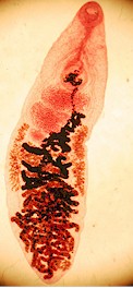
Anatomy of Dicrocoelium spp
Adult Dicrocoelium have the flat body and the oval shape typical for most flukes. They are smaller than liver fluke, ~10 mm long and ~2 mm wide. Their body is translucent. They have 2 prominent suckers, the oral sucker and the ventral sucker, both on the anterior part. They don't have cuticular spines. As other flukes they have no external signs of segmentation. The mouth ends in the pharynx, a muscular tube that allows sucking. The digestive system is blind (i.e. without anus: the only opening is the mouth) and not linear, as in most animals, but branched, ending in several blind ducts (called coeca). As most flukes, lancet flukes are simultaneous hermaphrodites, i.e. they have both male and female reproductive organs.
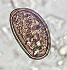
The eggs have an oval shape and are quite small (~25x40 micrometers), have a brownish color and contain a miracidium larva.
Life cycle and biology of Dicrocoelium spp
 Lancet flukes have an indirect life cycle with two intermediate hosts, a snail and an ant. It is one of the most conspicuous life cyclesamong the parasitic helminths.
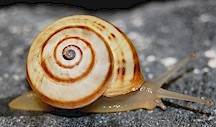
The eggs shed by adult flukes reach the host's gut with the bile and are expelled with the feces. Once outside the host terrestrialsnails ingest the eggs. Several species can act as intermediate hosts, depending on the regions and the environment, e.g. Theba spp, Zebrina spp, or Cionella spp for Dicrocoelium dendriticum; Limicolaria spp or Achatina spp for Dicrocoelium hospes. Inside the snails young miracidia hatch out of the eggs, penetrate the intestinal wall and develop to sporocysts, which on their turn multiply asexually, each one producing up to 100 daughter sporocysts. Each daughter sporocyst can produce up to 60 cercariae in the 3-4 months that it remains inside the snail. The snail encysts these cercariae and expels them in the form of sticky slime balls that adhere to the vegetation. Each slime ball can contain up to 100 cercariae.
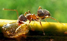
 Several ant species can act as the second intermediate host, e.g. Formica spp or Lasius spp for Dicrocoelium dendriticum; Dorylus spp or Cematogaster spp for Dicrocoelium hospes. These ants eat the slime balls. Inside the ants, most cercariae continue their development to metacercariae that are infective for the final host (cattle, sheep, goats, humans, etc.). These cercariae remain in the hemocoele of the ant. But one cercaria migrates to the sub-esophageal ganglion (a kind of brain) of the ant and manipulates its behavior by acting on the nerve cells. Towards the evening, instead of following the rest of the ant colony, the manipulated ant climbs on top of a blade of grass and remains there the whole night through until dawn. This increases the chance that a final host swallows it during early morning grazing. If a final host does not ingest the infected ant, it climbs down the blade of grass, joins the other ants and behaves normally until the next evening, when the process starts again. Otherwise, if it would remain the whole day on top of the blade of grass it would dry out and die exposed to excessive sun irradiation.
Once a final host ingests the infected ant while grazing, the metacercariae are released in the gut of the host and migrate to the liver through the common bile duct (ductus choledocus), i.e. they do not migrate through the liver tissues as Fasciola flukes do. Metacercariae complete the development to adults and start producing eggs in 8 to 12 weeks (= prepatent period) after being ingested. The adult flukes feed on bile and not on liver tissues.
Harm caused by Dicrocoelium infections, symptoms and diagnosis
 Most infections withDicrocoelium cause no symptoms or only slight ones. However, in case of heavy infestations (up to 50,000 flukes in one animal) the bile ducts can be irritated and become distended. Chronic infestations can turn into cirrhosis. Edema and blood loss(anemia) can also develop.
Nevertheless, infections with Dicrocoelium no are not as harmful as those with Fasciola hepatica or Fasciola gigantica and fatalities are rare. Economic damage is mostly due to condemnation of the livers at slaughter and to reduced productivity of affected livestock.
Diagnosis is done by egg detection in the feces or by identification of the flukes after necropsy. However, since the eggs are passed to the intestine only when the gall bladder is emptied, a negative fecal egg count is not conclusive, i.e. there can be false negatives.
Post a Comment